
In 1953, it was discovered that DNA was arranged in a double helix. Only 50 years later, the entire human genome was sequenced, all 3 billion base pairs. Now it is 2016, and we are in the post-human genome sequencing project era. It has been a little over a decade since the genome was fully sequenced, and scientists have been able to identify and classify DNA, discover how genes are regulated, identify how DNA is copied and passed on to daughter cells, link genes to certain diseases, but there are still so many questions that remain to be answered.
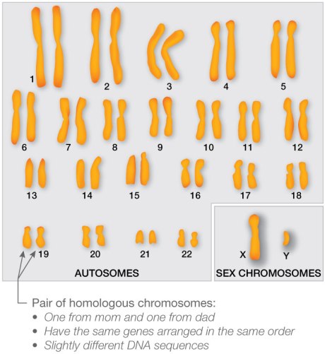
DNA is the language of our cells, except it has four letters (A, T, G, C) instead of the 26 letters in our alphabet. Those 4 letters combine in different ways to give your body instructions on how to function. Thanks to those 4 letters, you have a functioning heart, stomach, ball and socket joints, you name it. DNA is arranged in 23 chromosomes, kind of like a 23 volume encyclopedia (for an awesome supplement, click here, source of the karyotype below).
You would think that each chromosome codes for a group of related genes, yet cooperating genes are scattered all throughout. DNA is advertised as the genetic code/recipe/blueprint that provides instructions to make protein. To help illustrate this to high school students, I developed the activity below because it provides a meaningful opportunity to illustrate the concept that in order to make protein, genes are copied (transcription) and interpreted by RNA (translated).
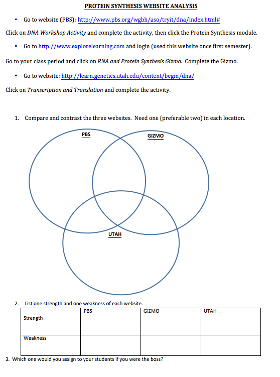
*Explorelearning.com has a fee and it might not be provided by your school *
I assign it for homework the day before we discuss protein synthesis in detail. The website involves interactive protein building on a very basic level. The Venn diagram section, the compare and contrast, is a great analysis activity that reinforces the concept of transcription and translation and forces students to process at that analysis level. The last portion involving identifying strengths and weaknesses is at the evaluation level, the mecca of learning. Working up the Bloom’s taxonomy ladder like this is critical scaffolding that builds a strong foundation in order to hang details on, making content retention easier.
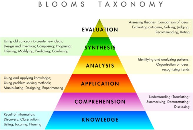
*Check out this wordpress blog here, the source of this picture. It does a great job explaining that the knowledge level questions teachers spend their time asking in class are less effective at content retention than high levels*
The warm-up the next day also scaffolds. At the beginning of class students discuss their answers to review the concept. At this point, even the ones that didn’t do the assignment (a sad reality of high school) have the opportunity to ask questions when their peers explain it in “kid” language (comprehension). Then, as a large group, I quickly click through each website and ask groups to present what they liked and what they didn’t (analysis). After about ten minutes, then they are primed and ready to hear all the details that are up ahead.
Once students have achieved mastery of protein synthesis, AP level classes will then go on to rock student’s world and tell them that protein is actually only 1.5% of the genome (the exon portion of the pie – see image from Campbell Biology, 9th edition, figure 21.7).
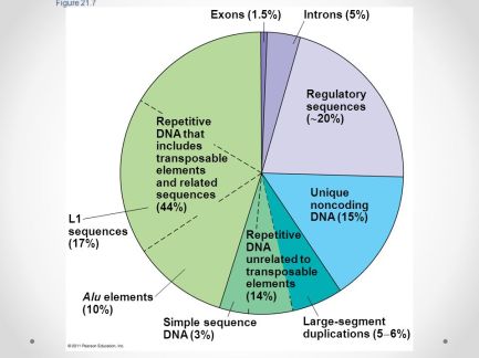
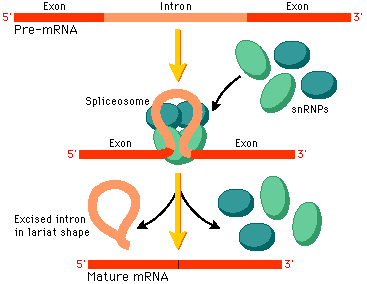
The fact that only 1.5% of all the 3 billion base pairs make protein is astounding. What about the rest of it? The introns (5%) are scattered within the coding portions of our genes (see picture from this website). In eukaryotes, genes have “alternative splicing”, meaning one gene can code for several different proteins by rearranging the location of the exons and introns within its coding sequence. For example, the image below has pre-mRNA for one of its protein. That same sequence might have different exons and introns arrangements, making an entirely different protein.
Regulatory sequences (20%) include promoters, which are regions that recruit transcription initiation, allowing the gene to be copied. An entire complex with many specific and non-specific proteins binds at a region upstream of the gene, and it requires the presence of many interacting proteins.
The unique noncoding DNA (15%) includes pseudogenes, or nonfunctional sequences that used to be genes. Repetitive DNA unrelated to transposable elements (14%) include centromeres and telomeres (see image taken from this website).
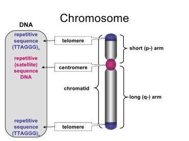
Telomeres are like the protective plastic at the end of your shoelaces (those actually have a word, and they are called aglets). At every division, a the telomeres shorten and shorten, and after about 50-100ish divisions, when the telomeres are no longer present, the cell commits suicide (cell suicide is called apoptosis). Seems like a tragic ending, but let’s face it, once the aglet comes off of the shoelace they go in the trash because the whole thing will unravel and be nonfunctional. The same phenomenon would happen with DNA. If the cell continued to divide, important genes would be destroyed, completely eliminating machinery for critical biological processes. Also, it is believed that this apoptosis prevents cancer, as with each division, more mutations accumulate, and any change in DNA has the potential to be deadly.
A whopping 44% of the human genome contains transposons, the wandering genes I mentioned in my last post. Some are believed to be silenced, most likely by their location in tightly coiled heterochromatin. Some are associated with disease . Others appear to be regulated by a special type of RNA (also coded by DNA) called siRNA, which binds to and inhibits the transposons activity. Transposons baffle me – with 44% of our genome mobile, how is there not massive mayhem? Genes would be disrupted and become nonfunctional if all 44% moved randomly throughout their lifetime. Surely, there has to be some sort of recognition site, or maybe the siRNA and its family members have a more significant role in silencing that is believed.
The amount of genetic information that we do not know about seems infinite, yet progress is still made and drug therapy is still developed that effectively influences our biology. The very beginning of this impossibly complicated quest involving DNA all started with the discovery of the double helix. Although Watson, Crick, and Wilkins were the Nobel Prize winners for this discovery, a woman named Rosalind Franklin provided the evidence that led to their epiphany. Her famous photograph 51, shown below, was stolen from her lab by Wilkins, who allegedly took it to Watson and Crick, and her picture is ultimately the reason the three men received the Nobel Prize 9 years later.
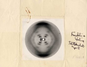
She did not achieve the Nobel Prize because she had passed away, most likely due to the intense x-ray work she used to study DNA (see the x-ray diffraction photograph 51 above). Not only did she not receive the Nobel prize, Watson wrote a book called The Double Helix in 1968 and ruthlessly criticized her. To see a fascinating movie about the whole saga, click here. Unlike my other clips, this one is an hour long movie. I was enraged on her behalf when I first saw it, and then proceeded to go home and show my entire family so that they would know this important story, one I think the whole world should be told.
Next post, I will focus on RNA. It is bothering me that the genomic pie chart above does not show the portion of DNA that is responsible for RNA synthesis, as there are at least seven RNAs (mRNA, tRNA, rRNA, siRNA, miRNA, piRNA, snRNP) that are functional and critical. There isn’t a lot of hype about the RNA, but it is just as critical a piece of the puzzle.

